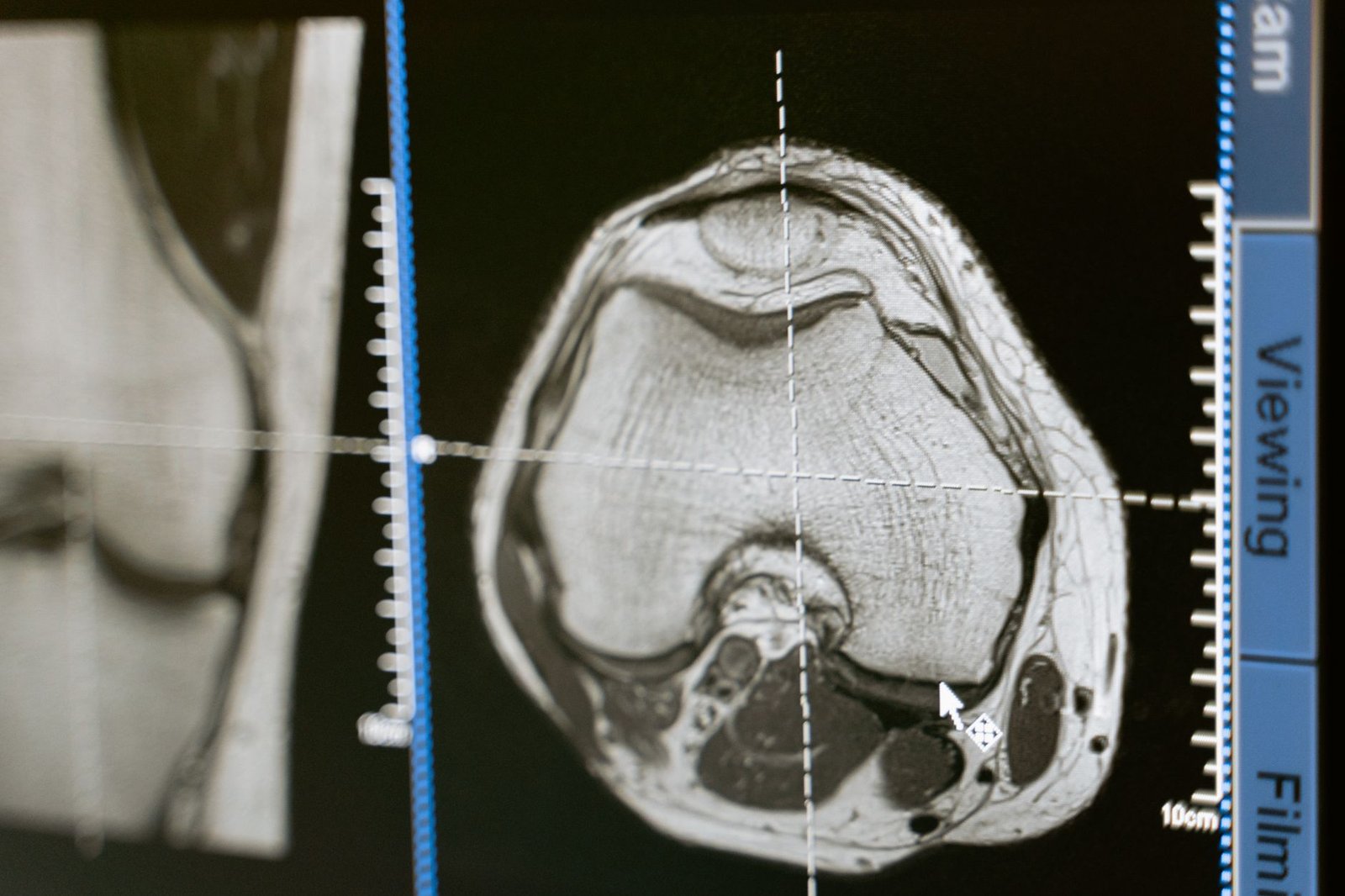
WHEN THE IMAGING DOES NOT MATCH THE PATIENT
There is a particular silence that follows a normal report. Nobody says you are making it up. The file simply stops moving, and you are left holding a study that answered a question you were not asking.
What the study was ordered to do
Imaging after a collision is ordered to exclude catastrophe: fracture, dislocation, cord compression, bleeding, a surgical emergency. It is very good at that, and a negative result is genuinely good news. It is a different instrument from the one that identifies which small, densely innervated structure is generating pain six months later, and it was never claimed to be that instrument by the radiologist who read it.
Two errors that look opposite and are the same error
Both come from treating a picture as a diagnosis.
- The normal scan, blamed on the patient. Nothing shows, therefore nothing is there, therefore this is deconditioning, or anxiety, or something to do with a claim. It is a conclusion drawn from the limits of a tool.
- The abnormal scan, blamed for the injury. Something shows — disc degeneration, a bulge, a cuff tear — and it becomes the explanation for everything. Findings that common are usually silent.
The prevalence data on the second error is not subtle. A systematic review of 3,110 asymptomatic people across 33 studies found disc degeneration in 37% of 20-year-olds rising to 96% of 80-year-olds, and disc bulges in 30% to 84% over the same span. In the Chingford general population cohort of 1,003 women aged 64 to 87, full-thickness rotator cuff tears were present in 22.2%, and 48.4% of those tears caused no symptoms at all.
A finding present in most pain-free people cannot, on its own, explain one person's pain. It also cannot date it, which is the use it most often gets put to after a collision.
The structures that are diagnosed by response, not by picture
Several of the most common sources of lasting pain after an injury have no imaging signature worth the name. They are identified by anesthetizing them and watching what happens.
- Cervical facet joints. In chronic neck pain after whiplash, controlled diagnostic blocks identified a painful facet joint in 54% of 50 consecutive patients, and placebo-controlled blocks put the overall prevalence at 60% in 68 consecutive referrals. Detailed on the whiplash page.
- Lumbar facet and sacroiliac joints. Same logic, same discipline, described on the facet page and the sacroiliac page.
- The disc that hurts versus the disc that looks bad. A separate problem with its own page: a painful disc and an ugly disc are not the same disc.
What a dynamic examination adds that a static image cannot
A magnetic resonance study images a limb lying still. Ultrasound can watch a tendon move, load a joint and observe it, and compare the injured side against the other one in real time. It shows the tendon that subluxes only at end range and the ligament that gapes only under stress — findings that are invisible when the part is at rest inside a magnet.
Guidance also decides whether treatment reaches its target. Systematic reviews and meta-analyses found ultrasound-guided injections more accurate than landmark-guided injections at the hip, and both more accurate and more effective at the shoulder girdle. An injection that did not reach the structure is not a treatment that failed. It is a treatment that never happened, and it is one of the reasons published results in this field vary as much as they do — the wider version of that argument is on why the trials disagree.
What we write down instead
A record that says “MRI unremarkable, continue therapy” carries nothing. The record we build states the mechanism and the date, the structures examined and how, the specific provocation and relief findings, what was injected including its preparation, how placement was confirmed, and function measured the same way at each visit so the numbers are comparable. Reporting standards in this field are poor enough that a systematic review found randomized trials of platelet-rich plasma for knee osteoarthritis adhere badly to the MIBO reporting guideline — if published trials do not characterize what they injected, a clinic note certainly will not unless someone decides it must.
The same record carries the terrain: the inflammatory and metabolic markers measured at baseline and again later. Not as background detail, but because they are the conditions the repair had to run in, and a result read without them is a result nobody can learn from. What we measure is set out separately.
Questions about imaging after an injury
If my MRI is normal, what is causing the pain?
Most often a structure the study is not built to show: a joint capsule, a ligament under load, a small nerve, or a sensitized pain system. In the neck specifically the most common identified source is a facet joint, which is set out on the whiplash page.
Should I get a better scan?
Sometimes, and more often the answer is a different kind of examination rather than a higher field strength — a dynamic ultrasound assessment, or a diagnostic block that tests a specific structure directly. What an assessment here involves is on the what-to-expect page.
The report mentions degeneration. Does that mean my injury did not cause this?
It means a common age-related finding is present, which is true of most people without pain — 96% of asymptomatic 80-year-olds have disc degeneration. A degenerate structure has less reserve and an event can exceed what remained. What that means for a joint that was injured rather than worn out is on the post-traumatic knee page.
Does ultrasound guidance actually change the result of an injection?
At the hip and the shoulder girdle the pooled evidence says yes on accuracy, and at the shoulder on effectiveness as well. Why we treat placement and preparation as part of the treatment rather than as technique is on the preparation page.
Nobody has been able to tell me what is wrong. Where do I start?
With a mechanism, a date and an examination aimed at candidate structures rather than at the existing report — then a diagnostic step that tests the leading candidate. That sequence is described on the tissue timeline page.
The diagnostic thread
- The neck injury a scanner cannot see
- A disc that hurts and a disc that looks bad
- Why the trials disagree
- The tissue clock after an injury
- The shoulder after a collision
- Why we ask for bloodwork
Bring the normal scan. It is still useful.
It tells us what has been excluded, which is where a real examination starts rather than where it stops.
12174 Natural Bridge Rd, Suite 303
St. Louis, MO 63044
Next to DePaul Hospital, just off the 270 and 70 junction, west of the airport.
Sources
- Brinjikji W, Luetmer PH, Comstock B, et al. Systematic literature review of imaging features of spinal degeneration in asymptomatic populations. AJNR Am J Neuroradiol, 2015. PubMed 25430861 doi:10.3174/ajnr.A4173
- Hinsley H, Ganderton C, Arden NK, Carr AJ. Prevalence of rotator cuff tendon tears and symptoms in a Chingford general population cohort, and the resultant impact on UK health services. BMJ Open, 2022. PubMed 36100305 doi:10.1136/bmjopen-2021-059175
- Barnsley L, Lord SM, Wallis BJ, Bogduk N. The prevalence of chronic cervical zygapophysial joint pain after whiplash. Spine (Phila Pa 1976), 1995. PubMed 7709275 doi:10.1097/00007632-199501000-00004
- Lord SM, Barnsley L, Wallis BJ, Bogduk N. Chronic cervical zygapophysial joint pain after whiplash. A placebo-controlled prevalence study. Spine (Phila Pa 1976), 1996. PubMed 8855458 doi:10.1097/00007632-199608010-00005
- Hoeber S, Aly AR, Ashworth N, Rajasekaran S. Ultrasound-guided hip joint injections are more accurate than landmark-guided injections: a systematic review and meta-analysis. Br J Sports Med, 2016. PubMed 26062955 doi:10.1136/bjsports-2014-094570
- Aly AR, Rajasekaran S, Ashworth N. Ultrasound-guided shoulder girdle injections are more accurate and more effective than landmark-guided injections: a systematic review and meta-analysis. Br J Sports Med, 2015. PubMed 25403682 doi:10.1136/bjsports-2014-093573
- Stone AV, Abed V, Owens M, et al. Randomized controlled trials on platelet-rich plasma for knee osteoarthritis poorly adhere to the Minimum Information for Studies Evaluating Biologics in Orthopaedics (MIBO) guidelines: a systematic review. Am J Sports Med, 2024. PubMed 38282598 doi:10.1177/03635465231185289